Boards reward rapid translation of anatomy into “illness scripts”—compact, testable stories that connect mechanism, key findings, and the next best step. Build your MSK core around three ladders: (1) motion unit (bone–joint–muscle–nerve), (2) load & failure (how tissue fails and how it presents), and (3) triage (red flags first). Start proximally and move distally. For each region, identify the prime movers, innervation, vascular supply, and stabilizers, then attach high-yield injuries and maneuvers. Example: the rotator cuff centers glenohumeral stability; supraspinatus initiates abduction (C5, suprascapular nerve). Painful arc and positive empty-can suggests supraspinatus pathology; weakness after fall on outstretched hand (FOOSH) favors tear—next best imaging often MRI if surgical planning is anticipated. As you study, convert each row into a flashcard that prompts for mechanism, exam maneuver, and disposition. Rehearse with time pressure: read the stem, identify the ladder (motion unit → load → triage), and pick the single next action that minimizes morbidity (immobilize, image, aspirate, or reassure). Localization collapses MSK neurology into a few anchors. Think “root → plexus → named nerve” and test both sensory territory and a single myotome movement. Cervical myotomes: C5 (shoulder abduction), C6 (elbow flexion/wrist extension), C7 (elbow extension/wrist flexion), C8 (finger flexion), T1 (interossei). Dermatomal pain down the arm or leg with reflex changes suggests radiculopathy; isolated motor/sensory deficits in a peripheral nerve territory suggests mononeuropathy. Boards love distinguishing C7 radiculopathy (triceps weakness, ↓ triceps reflex) from radial neuropathy (wrist/finger extension weakness, sensory dorsal hand). Exam stems often embed load or posture (crutches compressing axilla → posterior cord/radial issues). When localization is in doubt, “test one root and one nerve” approach: pick a myotome movement and a cutaneous patch that uniquely separates two contenders. Management emphasizes activity modification, splinting to protect function (e.g., wrist splint for radial palsy), and red flags (rapidly progressive weakness, sphincter signs → urgent imaging). Muscle/tendon and ligament injuries follow predictable mechanisms. Overhead activity provokes rotator cuff tendinopathy; axial load + valgus stress injures MCL; sudden deceleration with pivoting tears the ACL. Exam maneuvers pair with anatomy and are frequently the single best next step in the stem. On exam day, choose maneuvers that maximize separation between diagnoses (e.g., Lachman vs. anterior drawer). Early immobilization and protected weight bearing are low-risk decisions when severe injury is plausible; reserve advanced imaging for surgical planning or persistent deficits. Use MDSteps to convert what you just read into adaptive question blocks, missed-item flashcards, and readiness analytics. Understanding bone remodeling clarifies fractures and metabolic conditions. Osteoclast-mediated resorption and osteoblast formation are coupled by RANKL/OPG signaling. Pathology stems from imbalance: osteoporosis (reduced bone mass, normal mineralization), osteomalacia/rickets (defective mineralization, low vitamin D), and osteitis fibrosa (hyperparathyroidism). Boards test risk recognition (steroids, menopause, immobility) and first-line therapies: weight-bearing exercise, calcium/vitamin D, and antiresorptives (bisphosphonates) for appropriate T-scores or fracture history. For bone infection, osteomyelitis presents with fever, localized pain, elevated inflammatory markers; sickle cell disease favors Salmonella, otherwise Staph aureus. Early X-rays can be normal; MRI is sensitive. Management is early empiric antibiotics tailored to organism; surgical debridement for abscess or hardware infection. Don’t miss septic arthritis: hot, swollen joint, decreased ROM—aspirate before antibiotics when feasible; synovial WBC often >50,000 with neutrophil predominance. Fragility fractures (vertebral compression, hip) signal systemic disease. Use FRAX and DEXA-guided thresholds for therapy. In pediatrics, distinguish physeal (Salter-Harris) injury from normal variants; in elderly, low-energy hip fractures need prompt surgery and DVT prophylaxis. Tie bone biology back to management choices—for instance, why bisphosphonates reduce vertebral fractures (antiresorptive) yet atypical femur fracture risk rises with long-term use (oversuppressed remodeling). Rapid triage depends on pattern recognition. Inflammatory pain: morning stiffness >60 minutes, improves with activity, swollen/warm joints, high ESR/CRP; think RA, spondyloarthropathy, crystalline disease, infection. Mechanical pain: worsens with use, brief morning stiffness, focal tenderness (tendinopathy, OA, bursitis). Use distribution and time course to shortcut differentials: When stems force an immediate action, default to safety: aspirate the hot joint, immobilize suspected fracture/dislocation, and image when red flags exist. Use labs to confirm—not to delay—time-sensitive decisions. Analgesic choices hinge on mechanism and risk. For acute sprain/strain without red flags, start acetaminophen and NSAIDs; avoid opioids except for short-course, severe trauma scenarios. Add topical NSAIDs for localized OA. Glucocorticoids are potent but carry metabolic and infection risks; prefer local injections for focal inflammatory conditions (e.g., trigger finger, bursitis) after excluding infection and fracture. Neuropathic pain (radiculopathy) may respond to gabapentinoids or TCAs; weigh sedation and misuse potential. In inflammatory arthritis, short steroid tapers control flares while DMARDs (methotrexate first-line for RA) modify disease trajectory. Check pregnancy status, alcohol use, liver/kidney function, and give folate with methotrexate. Biologics target TNF, IL-6, or costimulation pathways; screen for TB and hepatitis B prior to initiation. Crystal disease flares are treated acutely with NSAIDs/colchicine/steroids; urate-lowering therapy (allopurinol) is preventive and continued during flares with prophylaxis. Exam logic: choose the safest effective regimen that aligns with the suspected mechanism and the patient’s comorbidity profile (CKD limits NSAIDs, anticoagulation complicates injections). Recognize drug–disease pitfalls (fluoroquinolones → tendinopathy; bisphosphonates → esophagitis if recumbent dosing). Convert MSK into durable skills through spaced retrieval and case-based practice. Use a repeatable weekly loop: (1) two focused Qbank blocks (mixed MSK + systems), (2) brief error autopsy mapping each miss to anatomy/illness-script ladders, (3) targeted Anki on mechanisms and maneuvers, (4) one mini-assessment with timed questions. Keep notes minimal and visual; prioritize tables and checklists you can rehearse under 60 seconds. The goal is fast pattern access, not encyclopedic recall. Use this list as your pre-exam warmup: if a stem triggers one of these, you likely know the correct “next step.”Anatomy → Function → Predictable Clinical Patterns
Structure Function Classic Presentation High-Yield Test Next Best Step Radial nerve (spiral groove) Wrist/finger extensors “Saturday night palsy,” wrist drop Sensory dorsum 1st web space; resisted extension Splint, avoid pressure; r/o humeral shaft fracture Ulnar nerve (cubital tunnel) Interossei, adductor pollicis Numb 4th/5th digits, intrinsic hand weakness Froment sign, Wartenberg sign Activity modification; consider nerve conduction Supraspinatus tendon Abduction initiation Lateral shoulder pain, night pain Empty-can, painful arc Activity + PT; MRI if trauma/weakness > surgical eval ACL Anterior tibial restraint Pop + swelling after pivot Lachman (> Anterior drawer) Brace + MRI; ortho referral Nerve Lesions, Dermatomes, and Myotomes—Fast Localization
Tendons, Ligaments, and Region-Based Maneuvers
Shoulder
Knee & Ankle
Turn this article into a smarter USMLE study plan
Practice the concept while it is fresh.
See how it works
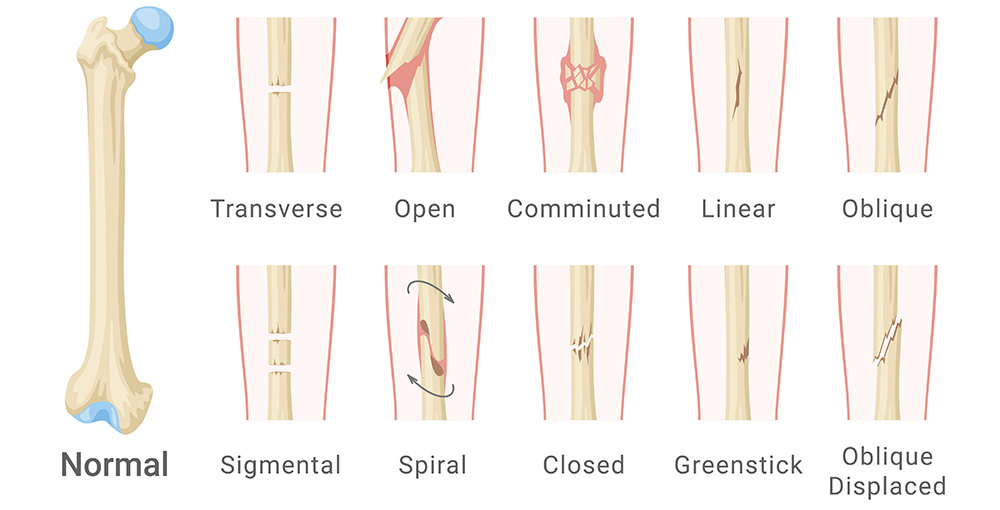
Bone Biology, Infection, and Metabolic Themes
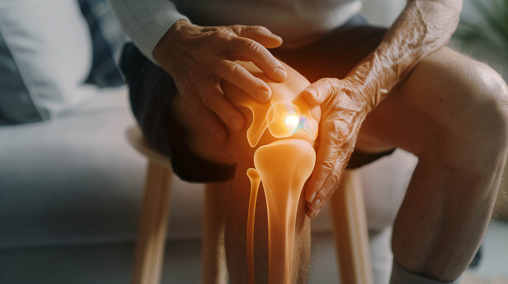
Inflammatory vs. Mechanical: Building Fast Illness Scripts
Pharmacology Across Steps: Analgesia, Steroids, and DMARD Logic
Practice Architecture: Qbanks, Retrieval, and a Weekly MSK Loop
Day Focus Primary Task Output Mon Upper limb 40 mixed Qs + review 3 cards: nerve, maneuver, dispo Wed Lower limb/spine 40 mixed Qs + review Flow for red flags vs. benign Fri Inflammatory vs mechanical 20 timed Qs Checklist: aspirate/image/immobilize Sun Imaging & emergencies Image identification drill Top 10 signs gallery Rapid-Review Checklist: Don’t Miss, Don’t Over-Treat
Sources & Further Study
Clinicals
Cross-Step MSK Mastery: From Anatomy to Illness Scripts
This guide links Step 1 structure/function with Step 2–3 patterns so MSK questions feel predictable on exam day—from nerve lesions and tendon tears to red-flag arthritis.
Full access: $27/mo
Lifetime: $299
16,000+ NBME-style questionsDepth-on-Demand™ explanationsCCS + readiness analytics
Monthly access. Cancel anytime. No free trial required.
Best next step