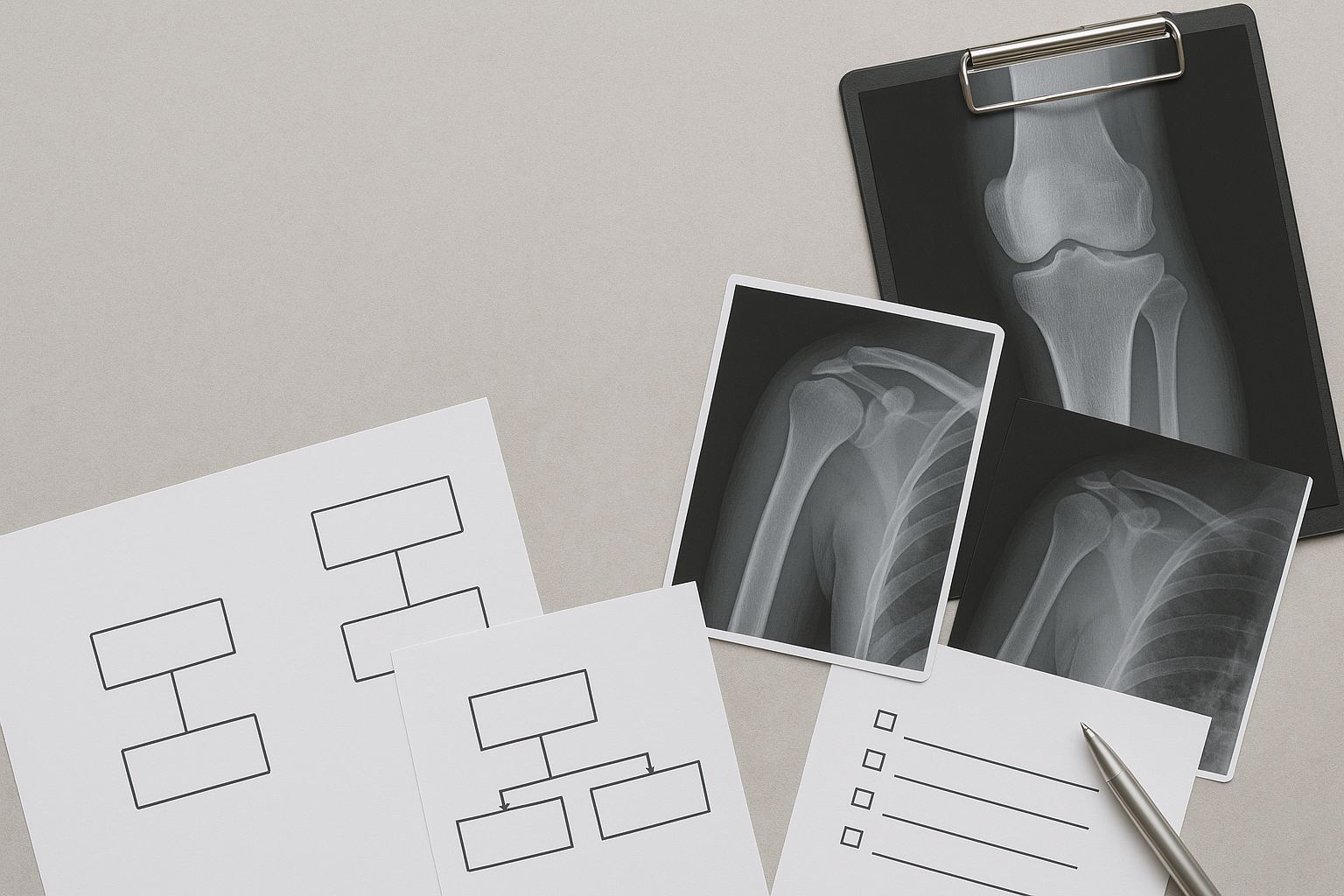
On Step 1, imaging is a reasoning task, not a radiology rotation. Your goal is to map clinical setting → suspected pathology → appropriate modality → signature phrase. Stems telegraph modality choices with age, time course, mechanism, exam sign, lab context, and specific adjectives. For fractures and dislocations in acute trauma, plain radiographs (XR) are usually first-line due to speed, availability, and low radiation. Computed tomography (CT) refines bony detail (e.g., complex intra-articular fractures, subtle vertebral injuries). Magnetic resonance imaging (MRI) excels at marrow, cartilage, ligaments, menisci, rotator cuff, and early osteomyelitis. Ultrasound (US) is clutch for dynamic tendon exams, effusions, and guiding procedures; in peds, it avoids radiation (e.g., hip effusion). Nuclear medicine (bone scan or tagged WBC) appears in stems emphasizing multifocality or occult stress injury when MRI is not mentioned. Translate descriptive cues quickly: “sunburst,” “Codman triangle,” and “metaphyseal lesion” cue aggressive bone tumors; “cortex-based lesion with night pain relieved by NSAIDs” suggests osteoid osteoma; “lucency at the scaphoid waist with snuffbox tenderness” implies occult scaphoid fracture (initial XR may be normal—immobilize and repeat imaging/MRI). Time language matters: acute (hours–days) pushes XR/US; subacute to chronic (weeks–months) pushes MRI/CT, depending on tissue. Infection keywords (“fever, elevated CRP, unremitting pain, post-op wound”) prime MRI for osteomyelitis or septic arthritis. Practice converting stems into this chain using MDSteps’ Adaptive QBank. Missed items automatically populate exportable flashcards—review the modality picked, the buzzword, and the pathophysiology; then re-test with spaced intervals to lock retrieval speed. Most trauma stems want XR first—because the question tests whether you recognize indications before escalating. Learn the classic XR views: shoulder (AP, axillary, scapular Y), elbow (AP/lateral with fat-pad sign), wrist (scaphoid view), knee (AP/lateral/sunrise), ankle (mortise). Beware normal-appearing XR in occult fractures: scaphoid, tibial stress, femoral neck, Lisfranc. The stem may emphasize persistent pain, focal tenderness, or high-risk location—these warrant MRI for marrow edema or CT for cortical detail. Stress fractures favor MRI (highest sensitivity early), while bone scans show increased uptake but lack specificity. Dislocations come with neurovascular pitfalls: anterior shoulder dislocation risks axillary nerve (deltoid numbness, weak abduction); posterior dislocation (seizure/electrocution) shows “light-bulb” sign; knee dislocation risks popliteal artery injury (check pulses, ABI, Doppler). Elbow fat-pad signs (“sail sign”) imply occult radial head fracture in adults and supracondylar fracture in children—CT/MRI is not always required on Step 1, but recognition is. For foot injuries, midfoot pain after twisting with plantar ecchymosis flags Lisfranc injury—XR weight-bearing views may reveal diastasis; CT further characterizes. Use MDSteps’ analytics dashboard to tag misses by “modality escalation” errors. Your study plan generator can then schedule targeted blocks on fracture patterns and neurovascular associations, accelerating pattern recognition before test day. In infectious MSK stems, the question is rarely “What does the MRI show?” It’s “Which modality confirms the suspicion fastest and changes management?” Fever, elevated CRP/ESR, refusal to bear weight (child), or a hot, swollen joint cues septic arthritis—ultrasound can detect an effusion at the bedside, but the diagnostic test is joint aspiration (cell count, Gram, culture). MRI of the limb/region evaluates osteomyelitis (bone marrow edema) and adjacent soft tissues; contrast enhances detection of abscess or sinus tracts, particularly in diabetic foot infection. Chronic or postoperative infection may present with subtle imaging changes; CT defines cortical sequestra, whereas MRI depicts marrow and soft-tissue involvement. Nuclear medicine can appear with language like “multifocal bone pain” or “history of prior malignancy ruled out”—bone scan is sensitive but nonspecific; tagged WBC scans increase specificity for infection. Beware “recent surgery” or “metal hardware”: MRI with metal artifact reduction or CT might be referenced; exam writers often emphasize clinical priority (debridement or aspiration) over fancy imaging. Drill this logic with MDSteps’ AI tutor: paste a tricky stem, ask “Which modality next and why?” and convert the explanation into a spaced flashcard. The automated deck export to Anki supports long-term retention of these high-yield patterns. MDSteps helps connect injury mechanism, pain location, exam maneuver, imaging threshold, and treatment decision inside realistic stems. Step 1 loves aggressive vs. benign descriptors. You won’t measure margins; you’ll map location + age + periosteal reaction. Aggressive periosteal patterns include sunburst and spiculated (osteosarcoma), onion-skin/lamellated (Ewing sarcoma), and Codman triangle (interrupted periosteum, often osteosarcoma or Ewing). Benign features include well-defined margins, sclerotic rim, and absence of cortical breakthrough (e.g., non-ossifying fibroma). Add the anatomic predilection and age: osteosarcoma → metaphysis of long bones in adolescents; Ewing → diaphysis in children/teens, systemic symptoms; chondrosarcoma → pelvis/proximal femur in adults. Classic benign-but-painful vignette: osteoid osteoma—adolescent with night pain relieved by NSAIDs; CT shows a small lucent nidus with surrounding sclerosis. Osteoblastoma is larger, often posterior elements of spine, less NSAID-responsive. Multiple myeloma presents with lytic “punched-out” lesions in older adults; skeletal survey or low-dose WB-CT is often mentioned. Fibrous dysplasia produces “ground-glass” matrix; enchondromas yield ring-and-arc calcifications in the hands. When malignancy is suspected, MRI defines marrow extent and soft-tissue mass; CT characterizes matrix mineralization. The stem might test awareness that biopsy is the definitive diagnostic step before definitive therapy. In your MDSteps QBank review, tag any tumor item by “age + location + periosteum.” The analytics dashboard will surface your error patterns (e.g., mixing osteosarcoma vs. Ewing), letting you drill with custom mini-blocks until recognition is instantaneous. Soft-tissue stems prioritize function over pixels. Rotator cuff: older patient with painful arc and weakness in abduction/external rotation—MRI confirms full-thickness supraspinatus tear; ultrasound is reasonable if availability or dynamic assessment is emphasized. Labral tears (throwers) often require MR arthrography in real life; on Step 1, they’re more likely to test special tests (O’Brien, apprehension) or that XR can be normal. Lateral epicondylitis is clinical; imaging is rarely needed. Knee: meniscal tear (twisting injury, joint line tenderness, locking) → MRI; ACL tear (pivot shift, immediate swelling) → MRI confirms; XR may show Segond fracture as a clue. Ankle: Achilles rupture (sudden pop, positive Thompson) is clinical; ultrasound visualizes tendon discontinuity. Plantar fasciitis is clinical; imaging if atypical. Muscle strain vs. tear: MRI grades severity; ultrasound can detect hematoma. Hemarthrosis after noncontact pivoting screams ACL and does not require emergent imaging unless red flags exist. Build speed: in MDSteps, filter practice for “sports medicine” tags and convert any shaky items into one-sentence flashcards (scenario → modality). Periodically run the Study Plan Generator to interleave MSK with neuro and cardio so retrieval stays robust. Pediatric bones are not small adult bones. Physis (growth plates) and more cartilage mean different injury patterns and imaging choices. Buckle (torus) fractures present after axial loading with cortical bulging—XR is diagnostic; management is simple immobilization. Greenstick fractures show cortical disruption on one side with bending on the other. Supracondylar humerus fractures (fall on outstretched hand) are common and dangerous—XR shows posterior fat pad; worry about brachial artery and median nerve. Toddler’s fracture (spiral tibia) can be subtle—XR may be initially normal; repeat imaging or MRI if symptoms persist. Nursemaid’s elbow (radial head subluxation) is clinical; imaging is unnecessary unless atypical or trauma is suspected. Hip disorders: transient synovitis vs. septic arthritis—both present with refusal to bear weight; fever and labs split them. Ultrasound detects effusion; aspiration rules in sepsis. Slipped capital femoral epiphysis (SCFE) in overweight adolescents presents with hip/knee pain and limited internal rotation; XR with frog-leg lateral shows epiphysis displacement (“ice cream slipping off cone”). Legg–Calvé–Perthes disease (idiopathic AVN of femoral head) appears in younger children; XR may be normal early; MRI shows AVN. Always consider radiation: US is favored for developmental dysplasia (DDH) before ossification; XR after 4–6 months. In MDSteps, tag pediatric MSK items with “physis/SCFE/effusion.” The platform’s analytics will surface if you over-order CT in kids—retrain with an ultrasound-first mindset for joints and radiation-sparing strategies elsewhere. Many stems hinge on recognizing complications, not the primary injury. Avascular necrosis (AVN) follows femoral neck fractures, dislocations, or steroid/alcohol exposure. XR can lag; MRI is most sensitive (subchondral signal changes, “crescent sign” later). Scaphoid proximal pole injuries carry high AVN risk due to retrograde blood supply—negative XR doesn’t exclude it; management is immobilization and advanced imaging if symptoms persist. Compartment syndrome is primarily clinical: pain out of proportion, pain with passive stretch, paresthesias, pulselessness (late). Imaging is not required for diagnosis; the question tests recognition and emergent fasciotomy. Fat embolism syndrome after long-bone fracture manifests with hypoxemia, neurologic changes, and petechiae; CXR may show diffuse infiltrates—supportive care is key. Osteomyelitis can complicate open fractures; stems that stress contamination, delayed closure, or exposed hardware should push you toward early antibiotics and debridement—MRI defines extent but never delays life- or limb-saving steps. Use MDSteps study blocks titled “Red Flags” to rehearse these triggers until they’re reflexive. The Adaptive QBank can interleave red-flag items across systems to avoid context traps. Here’s your rapid translator for common Step 1 imaging buzzwords. Don’t memorize in isolation—pair each keyword with age group, anatomic location, and clinical setting. Then rehearse retrieval by covering columns and recalling the pairings aloud or via MDSteps auto-flashcards. Turn this table into a drill: in MDSteps, star any question containing these phrases; the platform will auto-build a “Mini-Atlas” deck that you can export to Anki and rehearse in 2–3 minute micro-sessions. Timed blocks create tunnel vision. Use this three-pass approach to stay systematic. Pass 1 (10–20 seconds per stem): identify the clinical domain (trauma, tumor, infection, sports), age, and the most modality-linked buzzword. Ask: “Is XR adequate? Is soft tissue central (MRI/US)? Is there an aggressive periosteal pattern?” Eliminate options that mismatch domain (e.g., choosing CT for meniscal tear). Pass 2: commit to a modality and an expected finding. If the answer asks for management, ensure imaging aligns (e.g., aspiration before MRI in septic arthritis with effusion). Pass 3: sanity-check red flags (AVN risk, compartment syndrome, neurovascular injury) and confirm that your choice changes management. Work in sprints: Use MDSteps’ Study Plan Generator to schedule 40–60 question MSK sessions interleaved with neuro and renal. After each block, the analytics panel highlights error clusters (e.g., overusing CT, missing pediatric US). Convert each error into a concise flashcard: cue → dx → best modality → reason. Re-drill the deck on alternate days (spaced repetition) until your decision time drops below 15 seconds per imaging question. Finally, mix in 10-question “red flag” micro-blocks to keep complications salient. Use this on your final pass the week before the exam. Say each bullet aloud and visualize the modality and expected finding. Then run a mixed MSK set in MDSteps to verify speed and accuracy. Pro tip: in MDSteps, create a “Rapid Review – MSK Imaging” custom quiz. The Adaptive QBank will tune difficulty to your weak spots, while auto-flashcards keep the keyword→diagnosis links fresh through your final week.How to Read the Stem Like a Radiologist (Without Seeing the Image)
Clinical cue in stem Likely pathology Best initial modality Signature phrase to watch for High-energy trauma; deformity Fracture/dislocation XR (AP/lateral ± special views) “Intra-articular step-off,” “disruption of cortical continuity” Snuffbox tenderness after FOOSH Occult scaphoid fracture XR; if negative but high suspicion → MRI/CT “Proximal pole at risk of AVN” Insidious shoulder pain, overhead athlete Rotator cuff tear/tendinopathy MRI (or US if availability emphasized) “Full-thickness supraspinatus tear,” “subacromial impingement” Fever + joint pain; child Septic arthritis US (effusion) → urgent aspiration; MRI if deep joint “Joint effusion with synovial thickening” Night pain relieved by NSAIDs Osteoid osteoma CT (nidus); MRI may be less specific “Radiolucent nidus with surrounding sclerosis” Fractures, Dislocations, and Stress Injuries: XR First, Then Escalate
Escalation Algorithm (Boards-Style)
High-Risk Anatomical Sites
Infection vs. Inflammation: Osteomyelitis and Septic Arthritis in the Stem
Stem clues Likely diagnosis Imaging priority Action-triggering result Child with fever, refusing to bear weight; hip pain Septic arthritis (hip) US to detect effusion → aspiration Purulent fluid, high PMNs → urgent washout/abx Diabetic foot ulcer, swelling, foul drainage Osteomyelitis MRI with contrast preferred Marrow edema + abscess → surgical eval + abx Sickle cell disease, metaphyseal pain Osteomyelitis (Salmonella or S. aureus) MRI; bone scan if MRI unavailable Early marrow edema → start targeted abx MSK questions reward mechanism, exam clue, and next step—not guessing the label.
Still missing questions you thought you understood?
Bone Tumor Patterns: Decoding Periosteal Language and Location
Buzzword/age Likely entity Common site Go-to imaging clue Teen, painful metaphyseal mass; sunburst; Codman Osteosarcoma Distal femur, proximal tibia XR aggressive periosteal reaction; MRI for extent Child/teen, diaphyseal lesion; onion-skin; fever Ewing sarcoma Femur, pelvis XR lamellated periosteum; MRI marrow/soft tissue Night pain relieved by NSAIDs Osteoid osteoma Diaphysis cortex CT lucent nidus with sclerosis Older adult, lytic skull/spine Multiple myeloma Axial skeleton Survey XR/WB-CT “punched-out” lesions Soft Tissue and Sports Injuries: When MRI, When Ultrasound, When XR
Scenario Likely dx Best test XR role Painful arc, night pain, weakness ER Rotator cuff tear MRI (or US) Rule out calcific tendonitis/AC OA Twist + locking/catching Meniscal tear MRI Misses soft tissue; may show effusion Pivot + pop + rapid swelling ACL tear MRI Segond fracture clue Sudden calf pop; weak plantarflexion Achilles rupture US (dynamic) or MRI Usually normal Pearls
Pediatrics and Growth Plates: The Rules Change with Cartilage
Pediatric cue Likely diagnosis Imaging choice Why this test? Refusal to bear weight, afebrile Transient synovitis US (effusion), clinical follow Radiation-free; monitors effusion Fever + hip pain + limp Septic arthritis US → aspiration; MRI if deep Detects effusion; guides urgent management Overweight teen, hip/knee pain SCFE XR AP + frog-leg lateral Shows epiphyseal slip; urgent pinning Young child, insidious limp Perthes disease XR; MRI early AVN detection and staging Complications and Red Flags: AVN, Compartment Syndrome, Fat Embolism
Complication Flow Path (Decode the Stem)
Stem trigger Think of Imaging role Board-next step Hip dislocation/femoral neck fracture AVN MRI sensitive; XR late Urgent reduction/fixation; weight-bearing limits Severe limb pain + passive stretch pain Compartment syndrome None required Emergent fasciotomy (don’t delay) Long-bone fracture + hypoxemia + petechiae Fat embolism CXR diffuse infiltrates Supportive care; stabilize fractures Open fracture, contaminated wound Osteomyelitis risk MRI extent; XR for hardware Early abx + debridement Keyword → Diagnosis Mini-Atlas: Turn Buzzwords into Answers
Keyword in stem Likely dx Modality that shows it best Why it matters “Periosteal onion-skin” Ewing sarcoma XR for pattern; MRI for extent Aggressive tumor in diaphysis; systemic signs “Sunburst + Codman triangle” Osteosarcoma XR pattern; MRI staging Metaphyseal lesion in teens; chemo + resection “Ground-glass matrix” Fibrous dysplasia XR/CT Benign developmental; deformity risk “Lytic punched-out skull lesions” Multiple myeloma Survey XR or WB-CT Plasma cell malignancy; CRAB features “Nidus; pain relieved by NSAIDs” Osteoid osteoma CT Small cortical lucency; prostaglandin-driven pain “Fat-pad/sail sign (elbow)” Occult fracture XR lateral elbow Adults: radial head; kids: supracondylar “Snuffbox tenderness; normal XR” Scaphoid fracture MRI/CT Proximal AVN risk; immobilize “Crescent sign hip” AVN MRI early, XR later Subchondral collapse risk Putting It Together Under Timed Conditions: A 3-Pass Method
Rapid-Review Checklist + Exam-Day Essentials
Musculoskeletal
Imaging the MSK System for Step 1: What the Stem is Telling You
For MSK diagnosis and management
The mechanism usually tells you what failed.
Practice joint pain, injury mechanisms, back-pain red flags, rheum clues, exam maneuvers, imaging, and management decisions.
Full access includes Step 1, Step 2 CK, Step 3, CCS cases, analytics, auto-flashcards, and study planning.
Pivot-clue review
See the exact phrase in the stem that should have changed your decision.
Distractor trap logic
Learn why the answer you almost picked felt right—and why it was wrong for this patient right now.
Miss-pattern analytics
Turn repeated mistakes into targeted blocks, flashcards, and readiness signals.
Board-style takeaway: You’re not reading films—you’re translating stem language into the most likely modality, the expected finding, and the next-step decision. Use the tables and flow paths below to convert buzzwords (e.g., “periosteal reaction,” “night pain relieved by NSAIDs,” “snuffbox tenderness”) into rapid, test-ready answers. Tip: mirror your practice with the MDSteps Adaptive QBank and auto-generated flashcards to reinforce these patterns.
Score stuck after more questions?
Free reasoning diagnostic
Pivot clue isolatedDistractor trap explainedNext study target identified
No credit card required for the free reasoning review. Full access is $27/month after that. Cancel anytime.
Reasoning diagnostic
Still missing the same kinds of questions?
Read why that happens.
References & Further Reading