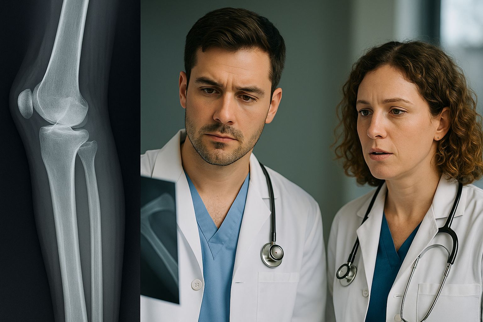
A Pattern-First Framework for MSK Pathology
MSK stems become predictable when you categorize them by four levers: age, anatomic site, tempo (acute vs. indolent), and a trigger phrase. Every item then reduces to a chain: clinical cue → core pathology process → hallmark finding → complication to anticipate. For example, “teen with painful metaphyseal mass; sunburst periosteal reaction” translates to an aggressive osteoid-producing tumor (osteosarcoma) that threatens local invasion and lung metastases. Similarly, “child with fever refusing to bear weight” triggers septic arthritis and a need for urgent aspiration before imaging delays care.
Begin each question by labeling the process as traumatic (fracture/dislocation; risk of AVN and compartment syndrome), infectious (osteomyelitis/septic arthritis), neoplastic (benign vs. malignant bone/soft-tissue), metabolic (osteoporosis/osteomalacia/Paget), or crystal/inflammatory (gout, pseudogout, spondyloarthropathies). Within each process, Step 1 frequently tests one pivotal decision (immobilize and escalate imaging; aspirate the joint; start antibiotics; recognize a red-flag complication) rather than nuanced subspecialty management. Pair this with modality logic—XR for fractures, MRI for marrow/soft tissue, CT for cortex and matrix characterization—to anchor the diagnostic step without overthinking.
Finally, train your eye for board buzzwords that carry mechanistic weight: “night pain relieved by NSAIDs” (prostaglandin-rich osteoid osteoma), “punched-out lytic lesions” (myeloma due to osteoclast activation), “onion-skin periosteal reaction” (Ewing’s small round blue cell tumor), “crescent sign” (subchondral collapse in AVN), and “calcifications in cartilage” (chondroid matrix). Use MDSteps’ automatic flashcards to couple each phrase with age and location, then interleave review using the platform’s study-plan generator. The goal is speed: convert stems into mechanisms within seconds, then choose the answer that prevents disability or death—exactly what exam writers reward.
Traumatic Pathology: Fractures, Dislocations, and Complications You Must Not Miss
Trauma questions hinge on two ideas: which injuries are occult but dangerous and which complications demand immediate action. Scaphoid fractures (snuffbox tenderness after FOOSH) may have initially normal radiographs; the proximal pole’s retrograde blood supply predisposes to avascular necrosis (AVN). Immobilize in a thumb-spica and escalate to MRI/CT if pain persists. Femoral neck fractures—especially displaced—threaten the retinacular vessels; AVN risk mandates urgent fixation. Talar neck injuries are similarly AVN-prone. Midfoot pain with plantar ecchymosis after twist suggests Lisfranc injury; failure to recognize diastasis causes chronic instability and arthritis.
Dislocations carry nerve-artery risks: anterior shoulder dislocation can damage the axillary nerve (deltoid numbness, weak abduction). Posterior shoulder dislocation appears after seizure/electrocution (arm internally rotated; “light-bulb” head). Knee dislocation risks the popliteal artery—check pulses, ABI, and use Doppler if abnormal; limb salvage depends on speed. Supracondylar humerus fractures in children threaten brachial artery and median nerve; look for anterior humeral line malalignment and fat-pad signs.
Board-Style Trauma Algorithm
- Stabilize ABCs; check neurovascular status before and after reduction.
- Obtain appropriate XR views; treat high-risk sites as fractures even if XR is negative.
- Immobilize; escalate to MRI for stress/marrow injury or CT for complex cortex.
- Address red flags immediately: compartment syndrome (pain out of proportion, passive stretch pain) → fasciotomy; open fractures → irrigation/debridement + antibiotics.
High-Yield Associations
- Scaphoid → AVN risk; do not “watch and wait” without immobilization.
- Femoral neck/talar neck → AVN; urgent fixation.
- Posterior hip dislocation → sciatic nerve injury; reduce early to protect head perfusion.
- Long-bone fracture + hypoxemia & petechiae → fat embolism (supportive care).
Use MDSteps analytics to tag “missed red flags.” The Adaptive QBank will resurface these patterns until your response is automatic.
Infectious Pathology: Osteomyelitis and Septic Arthritis by Setting
Infection stems test whether you recognize who gets infected, where the microbe enters, and what action changes outcome. Hematogenous osteomyelitis targets the metaphysis in children; Staphylococcus aureus is most common. Sickle cell disease adds Salmonella (and S. aureus). Diabetics with foot ulcers develop contiguous-spread osteomyelitis with polymicrobial flora, including anaerobes; probe-to-bone is a powerful bedside clue. IV drug use favors vertebral osteomyelitis and sternoclavicular infections. Postoperative infection language (“drainage, fever, new pain after fixation”) urges early debridement and culture.
Septic arthritis presents with an acutely hot, swollen, exquisitely tender joint and systemic symptoms. The diagnostic and therapeutic step is arthrocentesis—do not allow imaging to delay it. High synovial WBCs with PMN predominance and positive Gram or culture clinch the diagnosis. Neisseria gonorrhoeae dominates in sexually active young adults; think migratory polyarthralgia, tenosynovitis, and dermatitis. Prosthetic joints implicate biofilm-forming organisms (coagulase-negative staphylococci, S. aureus); management often includes hardware considerations.
| Clinical setting | Likely pathogen(s) | Clutch clue | Action-oriented step |
| Sickle cell disease | Salmonella, S. aureus | Metaphyseal pain, fever | MRI for early marrow edema; targeted antibiotics |
| Diabetic foot ulcer | Polymicrobial incl. anaerobes | Probe-to-bone positive | MRI with contrast; debridement + culture-guided therapy |
| IV drug use/back pain | S. aureus (incl. MRSA) | Vertebral tenderness; epidural abscess risk | MRI spine; emergent decompression if deficits |
| Young sexually active | N. gonorrhoeae | Triad: polyarthralgia, tenosynovitis, dermatitis | Arthrocentesis + empiric coverage while cultures pending |
Convert these into one-liners inside MDSteps auto-flashcards (setting → bug → must-do) and review with spaced repetition to keep pathogens sticky.
Master your USMLE prep with MDSteps.
Practice exactly how you’ll be tested—adaptive QBank, live CCS, and clarity from your data.
What you get
- Adaptive QBank with rationales that teach
- CCS cases with live vitals & scoring
- Progress dashboard with readiness signals
No Commitments • Free Trial • Cancel Anytime
Create your account
Bone Tumors: Age–Site–Matrix Rules That Win Points
Bone tumor items reward linking age, location, and matrix with periosteal reaction. Adolescents with metaphyseal lesions that produce osteoid (cloud-like density), aggressive periosteal reactions (sunburst, Codman triangle), and pain often have osteosarcoma—classically distal femur or proximal tibia. In children/teens, diaphyseal lesions with “onion-skin” periosteal layering and systemic symptoms fit Ewing sarcoma (t(11;22)). Adults with cartilage-producing tumors (rings-and-arcs calcifications) suggest chondrosarcoma—pelvis, proximal femur, or shoulder girdle. The exam may ask for the next step (MRI for extent, biopsy before definitive therapy) or for the favored site of metastasis (lungs).
Benign patterns still show up: osteoid osteoma causes night pain relieved by NSAIDs; CT reveals a small lucent nidus with sclerotic rim. Osteoblastoma is larger, favors posterior spine elements, and is less NSAID-responsive. Non-ossifying fibroma is eccentric, cortically based, and well circumscribed in kids; most lesions are incidental. Enchondroma in the hand shows chondroid calcifications; multiple lesions (Ollier, Maffucci) carry malignant risk.
| Buzzword | Likely tumor | Typical age/site | Teaching pearl |
| Sunburst + Codman triangle | Osteosarcoma | Teens; metaphysis around knee | Osteoid matrix; lungs first for mets |
| Onion-skin periosteum | Ewing sarcoma | Kids/teens; diaphysis long bones | Small round blue cells; t(11;22) |
| Night pain relieved by NSAIDs | Osteoid osteoma | Adolescents; cortical diaphysis | CT shows nidus; prostaglandin-mediated pain |
| Rings-and-arcs calcifications | Chondrosarcoma | Adults; pelvis/proximal femur | Cartilage matrix; local pain/swelling |
| Punched-out lytic skull/spine | Multiple myeloma | Older adults; axial skeleton | CRAB features; monoclonal spike |
In MDSteps, filter for “tumor” tag and use the platform’s analytics to spot whether you confuse matrix types. Drill short sets until recognition is instantaneous.
Metabolic Bone Disease: Lab Triads and Radiologic Clues
Metabolic bone questions reward “lab triads” matched to classic imaging. Osteoporosis features normal Ca/PO4/PTH with decreased bone mass and microarchitectural deterioration; fragility fractures at hip/vertebra/wrist dominate. Osteomalacia/rickets results from defective mineralization due to vitamin D deficiency or resistance, chronic kidney/liver disease, or anticonvulsants; labs show low/normal Ca, low phosphorus, and elevated alkaline phosphatase; imaging reveals Looser zones and, in children, metaphyseal cupping and fraying. Primary hyperparathyroidism raises Ca and lowers phosphorus with subperiosteal bone resorption (radial aspects of middle phalanges), bone pain, and renal stones. Paget disease (disordered remodeling) yields very high alkaline phosphatase with normal Ca/PO4; imaging shows mixed lytic/sclerotic lesions, bone enlargement, bowing, and skull involvement with hearing loss.
On exam day, couple each condition with a “must-not-miss” complication: vertebral compression fractures in osteoporosis; hypocalcemic tetany after sudden vitamin D repletion in severe deficiency is a distractor—more commonly watch for bone pain and proximal weakness. Paget increases high-output cardiac failure risk (AV shunts) and osteosarcoma transformation in a minority, especially with rapidly enlarging painful lesions. Hyperparathyroidism promotes nephrolithiasis and osteitis fibrosa cystica (“brown tumors”).
| Condition | Ca | PO4 | Alk Phos | Signature imaging | Key risk/complication |
| Osteoporosis | Normal | Normal | Normal | Decreased bone density (DXA) | Hip/vertebral fractures |
| Osteomalacia/Rickets | Low/normal | Low | High | Looser zones; metaphyseal cupping | Bone pain, deformities |
| Primary hyperparathyroidism | High | Low | Normal/high | Subperiosteal resorption | Nephrolithiasis; osteitis fibrosa |
| Paget disease | Normal | Normal | Very high | Mixed lytic/sclerotic, bone enlargement | High-output failure; osteosarcoma (rare) |
Build a “lab-first” deck in MDSteps: prompt side lists numbers only (Ca/PO4/Alk Phos), answer side gives the disease. This forces pathway-level recall instead of word-matching.
Crystal and Immune-Mediated Joint Pathology: Fast Differentials
Gout involves monosodium urate crystals (needle-shaped, negatively birefringent) precipitating in cold, peripheral joints—classically the first MTP (podagra). Triggers include dehydration, diuretics, high purine load, and rapid cell turnover. Acute attacks present with intense inflammatory pain; chronic disease leads to tophi and erosions with overhanging edges. Pseudogout (CPPD) features rhomboid crystals with positive birefringence and chondrocalcinosis on imaging; it targets larger joints like the knee and wrist and may flare after illness or surgery. On test day, remember that joint aspiration is the definitive diagnostic step for both when septic arthritis is in the differential.
For immune-mediated disease, Step 1 emphasizes rheumatoid arthritis (symmetric small-joint inflammatory arthritis, morning stiffness >1 hour, RF/anti-CCP positivity) with pannus formation leading to marginal erosions and joint space narrowing. Extra-articular manifestations include nodules, ILD, and anemia of chronic disease. Spondyloarthropathies (HLA-B27)—ankylosing spondylitis, psoriatic arthritis, reactive arthritis—present with inflammatory back pain improved with activity, enthesitis, and asymmetric oligoarthritis; axial disease shows sacroiliitis and syndesmophytes. SLE can cause non-erosive arthritis and immune complex deposition; think malar rash, cytopenias, and renal involvement.
| Entity | Defining clue | Key test | Board pitfall |
| Gout | Podagra; needle, − birefringence | Synovial crystal ID | Don’t confuse with septic joint—aspirate if febrile |
| Pseudogout (CPPD) | Knee/wrist; chondrocalcinosis | Rhomboid crystals, + birefringence | Often post-operative or hyperparathyroid association |
| RA | Symmetric MCP/PIP; morning stiffness | Anti-CCP, erosions on XR | Spare DIP (contrast with OA) |
| Ankylosing spondylitis | Inflammatory back pain; young man | Sacroiliitis; ↑CRP/ESR | Restrictive lung disease from chest wall fusion |
Quick Rules
- Hot, single joint + fever → aspirate first (always exclude sepsis).
- Needles and negative vs. rhomboids and positive helps separate gout from CPPD in seconds.
- DIP involvement favors OA or psoriatic arthritis, not RA.
MDSteps can auto-generate flashcards from your misses (e.g., “CPPD linked to hyperparathyroidism”). Export to Anki for spaced review.
Pediatrics: Growth Plate Injuries, Osteochondroses, and Unique Infections
Children are not small adults; cartilage-rich skeletons create unique pathologies. Salter-Harris fractures involve the physis; types III and IV traverse the epiphysis/articular surface, risking growth disturbance—urgent orthopedic evaluation is common in stems. Toddler’s fracture (spiral tibia) may be radiographically subtle—persistent limp prompts repeat XR or MRI. Supracondylar fractures threaten brachial artery and median nerve; watch for anterior interosseous deficit (can’t make “OK” sign) and compartment syndrome.
Self-limited osteochondroses also show: Osgood–Schlatter (traction apophysitis of tibial tubercle in adolescents with jumping sports) presents with localized tenderness and swelling; management is activity modification and quadriceps stretching. Legg–Calvé–Perthes disease (idiopathic AVN of femoral head) occurs in boys 4–8 with limp and limited internal rotation; early XR may be normal, MRI is sensitive. Slipped capital femoral epiphysis (SCFE) affects overweight adolescents; XR frog-leg lateral shows posterior-inferior displacement of the epiphysis; prompt pinning prevents AVN and chondrolysis.
Infection patterns differ: hematogenous osteomyelitis homes to metaphyses due to sluggish blood flow; septic arthritis of the hip presents with fever, refusal to bear weight, and irritability, demanding ultrasound-guided aspiration and washout. Always consider non-accidental trauma if the history is inconsistent or if fractures are in various stages of healing.
Pediatric Pearls That Save Points
- Suspected SCFE → non-weight-bearing and urgent ortho referral.
- Septic hip in a child → aspirate before antibiotics if stable.
- Salter-Harris III/IV → joint involvement; risk of growth arrest.
- Greenstick/buckle fractures are common and often managed conservatively—recognize the XR patterns.
Practice pediatric-only blocks in MDSteps; the platform’s analytics will surface overuse of CT in children and reinforce ultrasound-first habits.
Soft-Tissue & Rhabdomyolysis: Recognizing Tissue-Level Injury
Soft-tissue pathology is about function, not just pictures. Rotator cuff tears produce weakness in abduction (supraspinatus) and external rotation (infraspinatus/teres minor), with painful arc and night pain; degenerative tears predominate in older adults, acute avulsions in younger athletes. Achilles tendon rupture presents with a sudden “pop,” difficulty plantarflexing, and a positive Thompson test. Meniscal tears cause joint-line tenderness and locking after a twist; ACL tears follow pivoting with rapid hemarthrosis. Although imaging clarifies, the exam emphasizes clinical pattern recognition and avoiding dangerous misses like compartment syndrome (pain out of proportion; pain with passive stretch; paresthesia; pulselessness late) where the diagnostic test is intracompartmental pressure measurement—but treatment (fasciotomy) should not be delayed.
Rhabdomyolysis stems feature muscle pain/weakness after crush injury, prolonged immobilization, seizures, or statin/drug toxicity; labs show markedly elevated CK and myoglobinuria (heme-positive dipstick with few RBCs). The deadly complication is acute kidney injury from myoglobin-induced tubular necrosis; management prioritizes aggressive fluids, urine alkalinization in select cases, and discontinuation of offending agents. Hyperkalemia and arrhythmias are immediate threats. On exam, anticipate cofounders like heat stroke and malignant hyperthermia; both funnel into the same renal risk.
Soft-Tissue “Do-Not-Miss” List
- Compartment syndrome → treat clinically; do not wait on imaging.
- Open tendon or high-grade tear with functional loss → urgent orthopedic evaluation.
- Rhabdomyolysis → fluids early; monitor K+, Ca2+, and renal function.
- Acute septic joint masquerading as crystal flare → aspiration.
MDSteps’ AI tutor can turn any miss into a micro-explanation card (mechanism → consequence), which you can drill daily for durable patterning.
Rapid-Review Checklist: 30 Board-Style Pearls
Say these aloud, cover the answer, and retrieve from memory. Then run a 20-question mixed MSK block in MDSteps to confirm speed and accuracy.
- Snuffbox tenderness with normal XR → treat as scaphoid fracture; AVN risk.
- Posterior shoulder dislocation after seizure → internally rotated arm; “light-bulb” head.
- Long-bone fracture + hypoxemia + petechiae → fat embolism; supportive care.
- Pain out of proportion + passive stretch pain → compartment syndrome; fasciotomy.
- Child with fever refusing to bear weight → septic arthritis until proven otherwise; aspirate.
- Diabetic foot ulcer probing to bone → osteomyelitis; MRI with contrast and debridement.
- Teen, metaphyseal lesion, sunburst/Codman → osteosarcoma; lungs for mets.
- Child/teen, diaphyseal lesion, onion-skin → Ewing sarcoma; t(11;22).
- Night pain relieved by NSAIDs → osteoid osteoma; CT shows nidus.
- Older adult with punched-out skull lesions → multiple myeloma; CRAB features.
- Osteoporosis labs are normal; risk is fragility fracture.
- Osteomalacia → low PO4, high Alk Phos; Looser zones.
- Paget → very high Alk Phos; mixed lytic/sclerotic; hearing loss.
- Primary hyperparathyroidism → high Ca, low PO4; subperiosteal resorption.
- Gout vs. CPPD → needle (−) vs. rhomboid (+); both require aspiration if febrile.
- RA spares DIPs; erosive, symmetric small joints; anti-CCP positive.
- Inflammatory back pain better with exercise → spondyloarthropathy; sacroiliitis.
- SCFE → overweight adolescent; frog-leg lateral confirms; urgent pinning.
- Perthes disease → younger child; MRI detects early AVN.
- Salter-Harris III/IV → joint surface; growth arrest risk.
- Achilles rupture → positive Thompson; urgent immobilization/repair decisions.
- Meniscal tear → locking/catching; joint-line tenderness.
- ACL tear → pivot, pop, fast swelling (hemarthrosis).
- Vertebral osteomyelitis in IVDU → MRI spine; treat S. aureus.
- Open fracture → immediate antibiotics + irrigation/debridement.
- Osteoid osteoma pain is prostaglandin-mediated (NSAID-responsive).
- Bone matrix: osteoid (cloudy) vs. chondroid (rings-and-arcs) helps tumor ID.
- Cartilage-heavy pediatric joints → prefer ultrasound when possible.
- AVN locations: femoral head, scaphoid proximal pole, talar neck.
- Never let imaging delay limb-saving or life-saving care.
Lock these in with MDSteps’ Rapid-Review mode and export the list as Anki cards for daily two-minute refreshers.
References & Further Reading
100+ new students last month.

