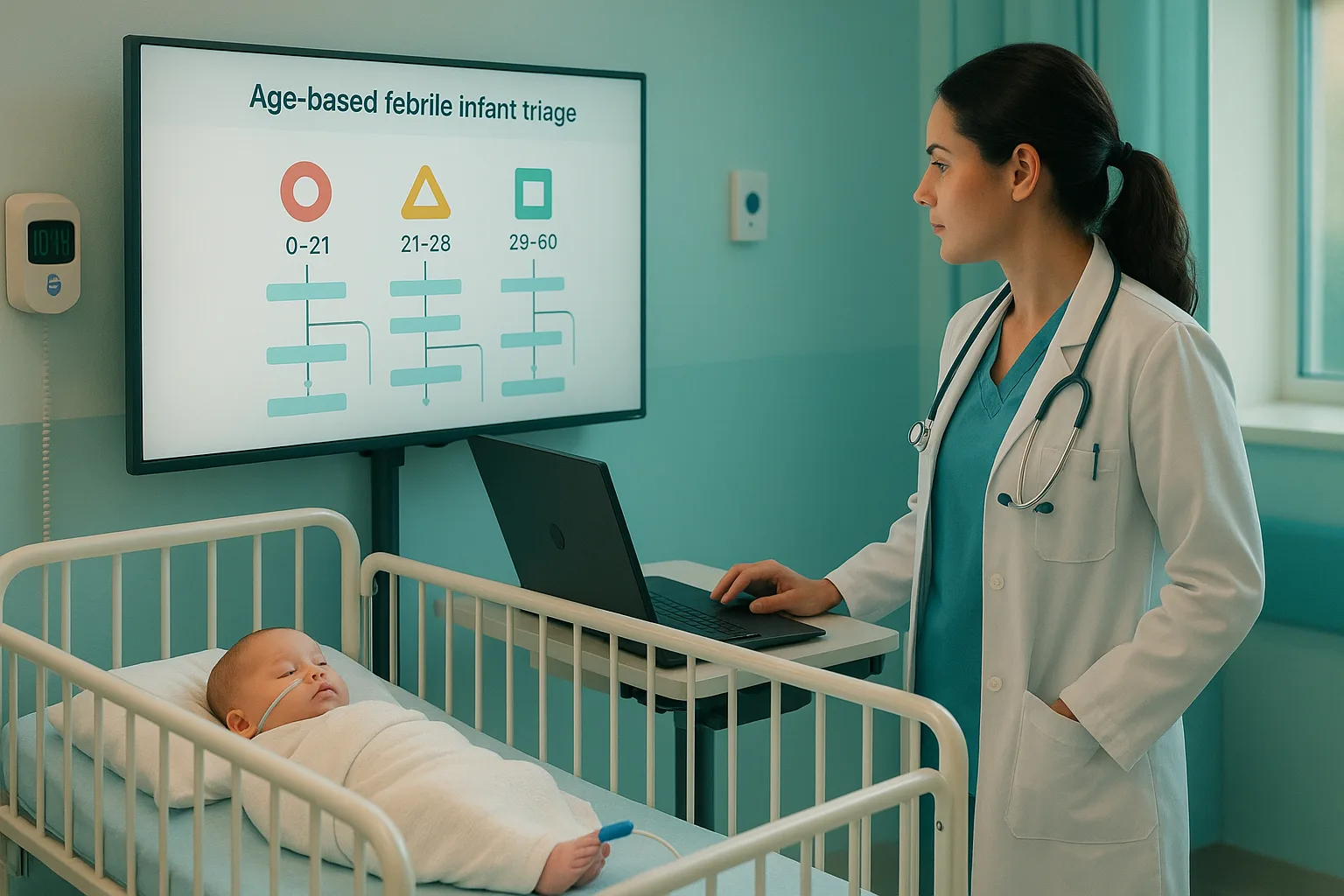
On Step 2 CK, you are expected to recognize dangerous bilirubin elevations, apply an evidence-based neonatal jaundice management algorithm, and know exactly when to start phototherapy or escalate to exchange transfusion. Neonatal jaundice is one of the most common issues in the nursery and a frequent topic on Step 2 CK. Up to two thirds of term newborns develop visible jaundice, but only a small fraction are at risk for bilirubin-induced neurologic dysfunction and kernicterus. Your job on the exam is to quickly separate benign physiologic patterns from pathologic hyperbilirubinemia that requires urgent treatment. That means thinking in hours of life, risk factors, and treatment thresholds rather than just memorizing random bilirubin numbers. Question writers exploit the fact that many students rely on intuition instead of a structured algorithm. They frame vignettes with subtle timing details, such as “jaundice noted at 18 hours” or “TSB 17 mg/dL at 60 hours in a 37-week infant,” and expect you to know when to simply follow-up, when to order labs, and when to start intensive phototherapy. The more you organize your approach as a standardized pathway, the more automatic these decisions become. In real practice, updated guidelines from the American Academy of Pediatrics (AAP) use hour-specific bilirubin thresholds for infants at or above 35 weeks’ gestation. These thresholds incorporate neurotoxicity risk factors (for example, hemolysis, sepsis, hypoalbuminemia, low gestational age) and are typically applied with digital tools such as BiliTool, PediTools, or local electronic medical record calculators. On test day, however, you must mentally approximate the same logic without clicking a calculator, so you should internalize a simplified but accurate framework. A high-yield way to prepare is to regularly practice “reading” bilirubin values in context: hours of life, gestational age, and presence of red flags. When you review pediatric questions in any QBank, look beyond whether the correct answer was “start phototherapy.” Ask yourself: Digital platforms such as the MDSteps Adaptive QBank can reinforce these mental habits by repeatedly surfacing bilirubin scenarios at different hours of life and difficulty levels. When your missed questions are automatically converted into flashcards, you start to see consistent patterns in which combinations of age, risk factors, and lab values trigger escalation of care. In this article, we will walk through the key physiology, define physiologic versus pathologic patterns, and then build a stepwise algorithm for managing jaundiced newborns. We will also highlight the kind of traps Step 2 CK writers love: benign breast milk jaundice versus red-flag early jaundice, inappropriate reassurance in hemolytic disease, or overuse of phototherapy in truly low-risk infants. By the end, you should be able to read a vignette and mentally run through a reproducible decision pathway within seconds. Before you can apply any algorithm, you need a firm grasp of why newborns become jaundiced in the first place. Neonates have increased bilirubin production due to high hematocrit and shorter red blood cell lifespan. At the same time, their hepatic conjugation capacity is limited because of low activity of UDP-glucuronosyltransferase. The result is an early-life imbalance that favors unconjugated bilirubin accumulation, particularly in the first week. The Step 2 CK exam loves to test distinctions between physiologic and pathologic jaundice. Physiologic jaundice usually: Pathologic jaundice is suggested by one or more of the following: Classically, unconjugated causes include hemolytic disease of the newborn (Rh incompatibility, ABO incompatibility, G6PD deficiency, hereditary spherocytosis), extravascular blood collections (cephalohematoma, bruising), polycythemia, and inadequate intake leading to increased enterohepatic circulation (sometimes labeled “breastfeeding jaundice” in the first week). Conjugated jaundice points you toward obstructive or hepatocellular disease such as biliary atresia, neonatal cholestasis, infections, or metabolic disorders. On Step 2 CK, a critical early branch in your mental algorithm is: Any “yes” here pushes you away from simple physiologic explanations and toward a more aggressive workup. Conversely, if the baby is term, feeding well, and becomes jaundiced at 48–72 hours with a modest bilirubin level, guidelines support conservative management as long as the value is clearly below treatment thresholds for that age and risk profile. Finally, remember the exam’s emphasis on bilirubin neurotoxicity. Unconjugated bilirubin can cross the immature blood–brain barrier and deposit in deep brain nuclei, producing acute bilirubin encephalopathy (hypotonia, lethargy, high-pitched cry, poor suck, progressing to hypertonia and seizures) and ultimately kernicterus with permanent motor and auditory deficits. The entire treatment pathway is designed to keep bilirubin well below levels that produce these outcomes, taking into account modifiers such as prematurity, hemolysis, acidosis, and sepsis. Examiners rarely give you a raw bilirubin number and ask “Is this high?” Instead, they test your ability to recognize patterns. Vignettes encode crucial information in age, maternal history, feeding status, and subtle exam findings. Your first task is to classify the scenario as physiologic, exaggerated physiologic, or clearly pathologic. Consider the following typical patterns: One common Step 2 CK trap is reassuring yourself that a baby is fine simply because the bilirubin is below 15 mg/dL. That is not safe reasoning. A value that is benign at 96 hours could be very concerning at 12–18 hours in a preterm infant with hemolysis. Always anchor your thinking in hours of life and gestational age, not just in absolute numbers. To keep these patterns organized, many students create a quick comparison table in their notes. A condensed version is shown below as a compact reference. Train next-best-step reasoning with adaptive clinical blocks, 50/50 trap analysis, and targeted review recommendations.
When studying, force yourself to label each vignette’s pattern explicitly after you review the answer
explanation. Over time, you will automatically recognize early-onset hemolysis versus benign late
breast milk cases. Platforms like MDSteps, which turn your misses into focused flashcards, are
particularly useful here because you repeatedly re-encounter similar scenarios until the pattern is
deeply ingrained.
Now that you can recognize common patterns, it is time to translate them into a structured decision pathway. While real-world clinicians use detailed nomograms, Step 2 CK expects a simplified schema that still respects guideline logic. A practical mental algorithm can be broken into four big steps: initial assessment, laboratory confirmation, risk stratification, and treatment versus monitoring.
The first branch in your algorithm answers three questions:
Any sick appearance (poor feeding, fever or hypothermia, respiratory distress, lethargy) should push
you toward sepsis evaluation alongside bilirubin assessment. Jaundice in the first 24 hours is
pathologic until proven otherwise and should immediately prompt total and direct bilirubin, blood type
testing, Coombs, complete blood count, and reticulocyte count. Do not “watch and wait” in these
scenarios on the exam.
In well-appearing infants with visible jaundice, transcutaneous bilirubin screening or a total serum
bilirubin level is obtained, followed by fractionation if indicated. The next step is to classify the
bilirubin as primarily unconjugated or conjugated. Conjugated hyperbilirubinemia should trigger a
cholestasis evaluation, abdominal ultrasound, and early specialist referral, not phototherapy.
Unconjugated patterns keep you within the classic neonatal algorithm.
Risk stratification integrates:
Conceptually, the more risk factors present, the lower your tolerance for a given bilirubin level at a
particular hour of life. Updated guidelines use specific curves for infants with and without
neurotoxicity risk factors. For Step 2 CK, internalize that a hemolytic, late-preterm infant with a
rapidly rising bilirubin should receive phototherapy at lower levels and be monitored closely, whereas
a robust term infant with no risk factors and a slowly rising bilirubin can be safely observed when
clearly below treatment lines.
Once you have a bilirubin value and a risk category, your choices narrow to three:
In exam questions, you are often asked for the next best step, not for every component of the
algorithm. Anchor your answer to where the infant sits relative to the appropriate curve for age and
risk, even if the nomogram itself is not shown.
The practical challenge on Step 2 CK is applying hour-specific thresholds without having the actual graph in front of you. You are not expected to memorize exact numbers, but you should know relative positions and principles. Several online tools implement guideline curves, but vignettes are written so that a clearly appropriate choice stands out when you understand the logic. A useful framing is to think about three ages: less than 24 hours, approximately 24–48 hours, and greater than 72 hours in term or near-term infants. In the first 24 hours, any visible jaundice is concerning and high values require urgent treatment. Between 24 and 48 hours, thresholds for phototherapy are relatively lower, and even mid-teens bilirubin levels can be worrisome in the presence of risk factors. After 72 hours, term infants can tolerate somewhat higher bilirubin levels before treatment is indicated, especially if they are otherwise thriving. Hemolysis, prematurity, and sepsis all shift these thresholds downward. For example, a late preterm infant with a positive direct antiglobulin test and rapid bilirubin rise should receive phototherapy at a lower level than an otherwise healthy term infant at the same hour of life. Vignettes often emphasize these risk factors explicitly, signaling that you should choose a more aggressive option. It helps to think qualitatively about distance from the phototherapy line. If the question stem tells you that the bilirubin is only slightly below what would require treatment, or that it is “within 2 mg/dL of the exchange transfusion threshold,” you should default toward initiating therapy and escalating care rather than discharging the infant. The goal is always to intervene before neurologic injury becomes likely. On exam questions, phototherapy is generally preferred over exchange transfusion when the bilirubin is at or modestly above the treatment threshold with no neurologic signs. Exchange transfusion is reserved for dangerous levels or for failure of intensive phototherapy with ongoing rise. Recognizing when a number has crossed that line is less about memorizing mg/dL cutoffs and more about integrating gestational age, risk factors, and rate of rise as the vignette presents them. Remember that phototherapy targets unconjugated bilirubin. If the infant has conjugated hyperbilirubinemia with acholic stools or dark urine, starting phototherapy misses the point and will not fix the underlying disease. The appropriate response is to evaluate for biliary atresia, metabolic disease, or other hepatobiliary pathology and involve subspecialists early. The most high-stakes branch of the algorithm is deciding when to go beyond standard phototherapy and escalate care. Exchange transfusion is an invasive procedure with its own risks, so guidelines reserve it for infants who are either at or above critical bilirubin levels, or who show signs of acute bilirubin encephalopathy despite intensive therapy. On Step 2 CK, red flags that should push you toward escalation include: In these scenarios, the correct answer often includes both immediate management and disposition: start or continue intensive phototherapy, initiate intravenous fluids, urgently consult neonatology, and transfer to a higher level of care for possible exchange transfusion. Exam writers test your ability to recognize that “watchful waiting” is unsafe when clinical signs suggest brain involvement or when bilirubin levels are dangerously close to critical thresholds. Another escalation decision point concerns early cholestatic disease such as biliary atresia. While unconjugated hyperbilirubinemia responds well to phototherapy, conjugated jaundice with acholic stools requires prompt diagnostic workup and surgical referral. Delayed recognition can permanently affect outcomes. If the vignette mentions persistent jaundice beyond two weeks with pale stools and dark urine, the “next best step” is not to repeat bilirubin levels indefinitely but to initiate evaluation for obstructive causes. When studying, focus on clinical reasoning rather than memorizing every detail of exchange transfusion protocols. Ask yourself: “In this vignette, is the risk of neurologic injury high enough that a conservative option would be irresponsible?” If yes, you should lean toward aggressive treatment choices in line with contemporary guidance. As you approach test day, your goal is not to recite guideline tables but to apply a structured approach under time pressure. A concise rapid-review checklist can keep your thinking organized when you see jaundice in a vignette.
As you review practice questions, consider building your own mini flowchart or flashcard that encodes
these key branches. The more you rehearse this logic, the faster and more confidently you will answer
bilirubin questions on Step 2 CK. When your QBank platform automatically tracks your performance and
highlights weak areas—such as late-preterm infants with hemolysis or breastfed babies with early
jaundice—you can systematically close those gaps before exam day.
With a clear mental pathway and repeated exposure to realistic vignettes, neonatal jaundice questions
become reliable points on your score sheet rather than anxiety triggers.
Medically reviewed by: Amara Singh, MD, FAAP – Board-Certified Pediatrician Why Neonatal Jaundice Matters for Step 2 CK and Clinical Practice
Understanding Bilirubin Physiology and Types of Neonatal Jaundice
Physiologic vs Pathologic Jaundice: Pattern Recognition on Step 2 CK
Turn this into Step 2 decision practice
Stop picking between two good-sounding answers.
See how it works
Pattern
Onset
Key Clues
Step 2 CK Implication
Physiologic
> 24 h (peak day 3–5)
Well infant, unconjugated, no risk factors
Usually observe, ensure follow-up
Breastfeeding jaundice
First week
Poor intake, weight loss, few diapers
Optimize feeds, sometimes supplement; treat if thresholds met
Breast milk jaundice
Day 5–7, persists
Thriving infant, high but stable levels
Continue breastfeeding; treat only if levels exceed thresholds
Hemolytic disease
< 24 h
Anemia, DAT+, maternal O or Rh−, G6PD, spherocytosis
Labs, early treatment, lower thresholds for phototherapy
Conjugated jaundice
Often after first week
Acholic stool, dark urine, hepatomegaly
Workup for cholestasis; phototherapy not effective
Building a Stepwise Clinical Algorithm for Newborn Bilirubin Elevations
Step 1: Initial assessment and red-flag screen
Step 2: Laboratory confirmation and classification
Step 3: Risk stratification and threshold comparison
Step 4: Decide between phototherapy, escalation, or observation
When to Start Phototherapy: Applying Guideline Thresholds Without a Nomogram
Escalation of Care: Exchange Transfusion, Red Flags, and Kernicterus Prevention
Rapid-Review Checklist and Exam-Day Strategy for Bilirubin Questions
Rapid-Review Checklist
Exam-Day Essentials
References
Algorithms & Diagnostics
Neonatal Jaundice Algorithm: How to Manage Bilirubin Elevations on Step 2 CK

Full access: $27/mo
Lifetime: $299
Clinical decision blocks50/50 trap analysisWeak-area review
Monthly access. Cancel anytime. No free trial required.
Step 2 reasoning